


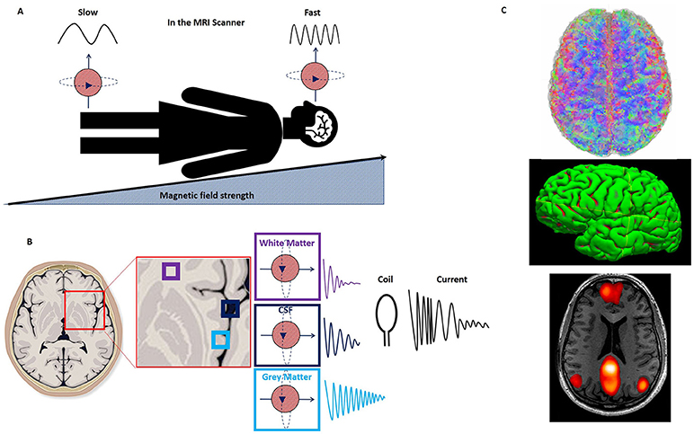
Magnetic Resonance Imaging (MRI) is a powerful diagnostic tool that relies on fundamental principles of physics to visualize internal structures of the human body in great detail. At its core, MRI technology is based on nuclear magnetic resonance—a phenomenon where atomic nuclei absorb and re-emit electromagnetic energy when placed in a magnetic field. Specifically, hydrogen nuclei (protons), abundant in the human body due to water content, are targeted by the MRI scanner. When exposed to a strong magnetic field, these protons align with the field. A radiofrequency pulse then temporarily knocks them out of alignment, and as they return to their original state, they emit signals that are used to construct detailed images.
The strength and precision of MRI come from its non-invasive approach and its ability to distinguish between different tissue types based on proton density and relaxation times. This precision is due to two key physical processes: T1 and T2 relaxation. These refer to the rates at which excited protons realign with the magnetic field and lose coherence with each other, respectively. Variations in these rates between tissues such as fat, muscle, and tumors help generate contrast in the resulting images. The technology behind MRI exemplifies how advanced physics principles are directly applied to improve modern medicine.
MRI physics continues to evolve, with ongoing research focusing on faster scan times, improved resolution, and reduced noise. Innovations like functional MRI (fMRI) allow scientists to map brain activity by detecting blood flow changes, a technique that opens doors in neuroscience and psychology. Understanding the physics behind MRI not only deepens appreciation for medical imaging but also highlights the critical role physics plays in technological and healthcare advancements.